

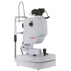
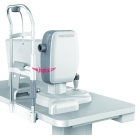
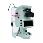
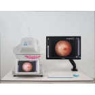
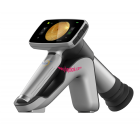
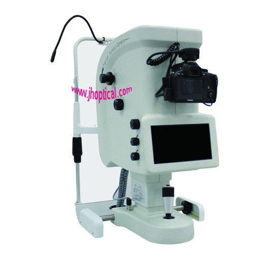
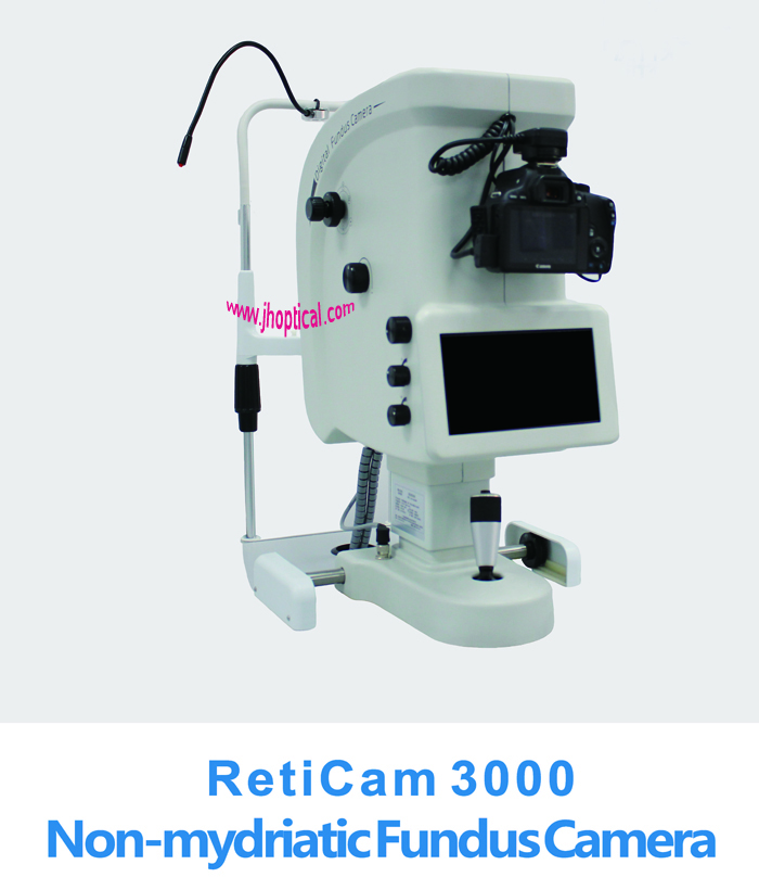
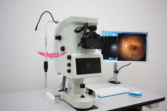
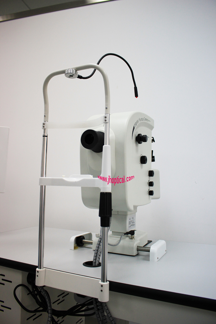
RetiCam 3000 Non-mydriatic Fundus Camera
Product Name:
RetiCam 3000 Non-mydriatic Fundus CameraModel No.:
RetiCam 3000Minimum Order:
1
Product Abstract:
Angle:42°/50° | Min pupil:3.3mm | 18 mega pixes | Diopter:±15D | Require external SRL
- Product Description
CE
RetiCam 3000 Non-mydriatic Fundus Camera
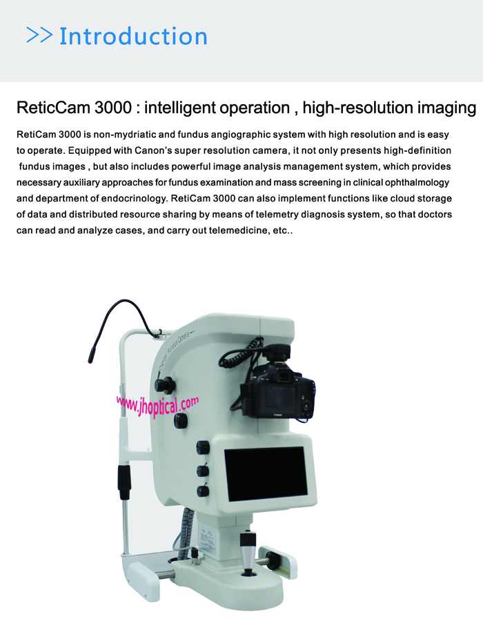
Introduction
ReticCam 3000 : intelligent operation , high-resolution imaging
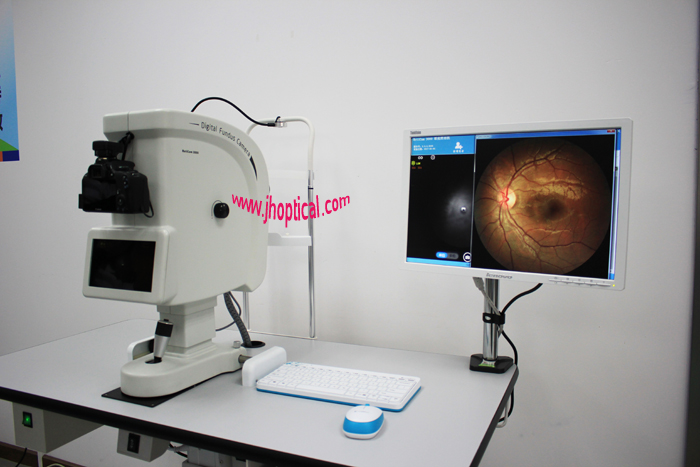
RetiCam 3000 is non-mydriatic and fundus angiographic system with high resolution and is easy to operate. Equipped with Canon’s super resolution camera, it not only presents high-definition fundus images , but also includes powerful image analysis management system, which provides necessary auxiliary approaches for fundus examination and mass screening in clinical ophthalmology and department of endocrinology. RetiCam 3000 can also implement functions like cloud storage of data and distributed resource sharing by means of telemetry diagnosis system, so that doctors can read and analyze cases, and carry out telemedicine, etc..Characteristics
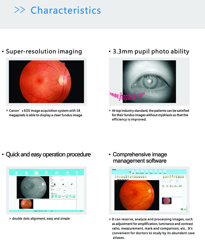
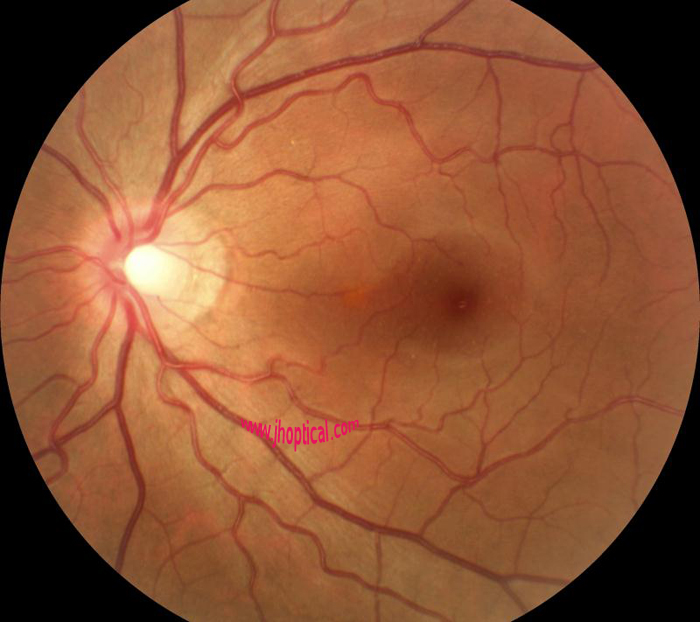
Super-resolution imaging
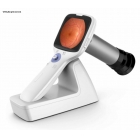
Canon’s EOS image acquisition system with 18 megapixels is able to display a clear fundus image
3.3mm pupil photo ability
At top industry standard, the patients can be satisfied for their fundus images without mydriasis so that the efficiency is improved.
Quick and easy operation procedure
Double dot anxiliary focus makes it easy to use.
Comprehensive image management software
It can reserve, analyze and processing images, such as adjustment for amplification, luminance and contrast ratio, measurement, mark and comparison, etc.. It's convenient for doctors to study by its abundant case atlases.Specifications:
Image Acquisition System
Acquisition mode: non-mydriatic/ mydriatic photograph; red free; eye’s anterior segment photography; fundus fluorescein angiography (optional)
Angle of vision: 42°(non-mysriatic)/50°
Working distance: 42mm
Requirement for pupil: ≥3.3mm
Digital acquisition form: external SRL
Sensor : 18 megapixels
Diopter compensation: ±15D
Anxiliary focus mode: double dot anxiliary focus
Others
Size:500 mm (L) x 195 mm (W) x 540 mm (H)
Weight: 20.3kg
Power supply: 220V/50Hz/5A
Note:with development of technology, specifications and design are subject to change without notice.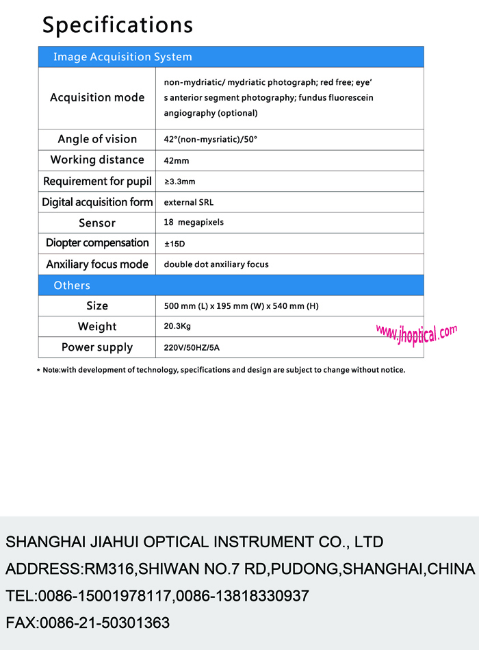
- Related Products
- [Return Home] [Print] [Go Back]
Contact Us
Contact:
Mr. Li YiyangTel:
+86 15001978117Fax:
+86-21-50301363E-mail:
jiahe@jhoptical.com-


