

Product Articles
- 2013-08-13 09:57:30
-
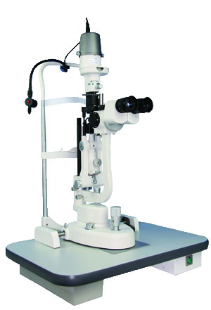
The slit lamp is an instrument consisting of a high-intensity light source that can be focused to shine a thin sheet of light into the eye. It is used in conjunction with a biomicroscope. The lamp facilitates an examination of the anterior segment, or frontal structures and posterior segment, of the human eye, which includes the eyelid, sclera, conjunctiva, iris, natural crystalline lens, and cornea. The binocular slit-lamp examination provides a stereoscopic magnified view of the eye structures in detail, enabling anatomical diagnoses to be made for a variety of eye conditions. A second, hand-held lens is used to examine the retina.
General procedure
While a patient is seated in the examination chair, they rest their chin and forehead on a support to steady the head. Using the biomicroscope, the ophthalmologist or optometrist then proceeds to examine the patient's eye. A fine strip of paper, stained with fluorescein, a fluorescent dye, may be touched to the side of the eye; this stains the tear film on the surface of the eye to aid examination. The dye is naturally rinsed out of the eye by tears.
A subsequent test may involve placing drops in the eye in order to dilate the pupils. The drops take about 15 to 20 minutes to work, after which the examination is repeated, allowing the back of the eye to be examined. Patients will experience some light sensitivity for a few hours after this exam, and the dilating drops may also cause increased pressure in the eye, leading to nausea and pain. Patients who experience serious symptoms are advised to seek medical attention immediately.
Adults need no special preparation for the test; however children may need some preparation, depending on age, previous experiences, and level of trust.
Variations in methods
Observation by optical sectionObservation with an optical section or direct focal illumination is the most frequently applied method of examination with the slit lamp. With this method, the axes of illuminating and viewing path intersect in the area of the anterior eye media to be examined, for example, the individual corneal layers.
Direct diffuse illuminationIf media, especially that of the cornea, are opaque, optical section images are often impossible depending on severity. In these cases, direct diffuse illumination may be used to advantage. For this, the slit is opened very wide and a diffuse, attenuated survey illumination is produced by inserting a ground glass screen or diffuser in the illuminating path.[11] "Wide beam" illumination is the only type that has the light source set wide open. Its main purpose is to illuminate as much of the eye and its adnexa at once for general observation.
Indirect illuminationWith this method, light enters the eye through a narrow to medium slit (2 to 4 mm) to one side of the area to be examined. The axes of illuminating and viewing path do not intersect at the point of image focus, to achieve this; the illuminating prism is decentered by rotating it about its vertical axis off the normal position. In this way, reflected, indirect light illuminates the area of the anterior chamber or cornea to be examined. The observed corneal area then lies between the incident light section through the cornea and the irradiated area of the iris. Observation is thus against a comparatively dark background.
Retro-illumination
In certain cases, illumination by optical section does not yield sufficient information or is impossible. This is the case, for example, when larger, extensive zones or spaces of the ocular media are opaque. Then the scattered light that is not very bright normally is absorbed. A similar situation arises when areas behind the crystalline lens are to be observed. In this case the observation beam must pass a number of interfaces that may reflect and attenuate the light.
Scattering sclero-corneal illumination
With this type of illumination, a wide light beam is directed onto the limbal region of the cornea at an extremely low angle of incidence and with a laterally de-centered illuminating prism. Adjustment must allow the light beam to transmit through the corneal parenchymal layers according to the principle of total reflection allowing the interface with the cornea to be brightly illuminated. The magnification should be selected so that the entire cornea can be seen at a glance
Fundus observation and gonioscopy with the slit lamp
Fundus (eye) observation is known by the ophthalmic and the use of fundus cameras. With the slit lamp, however, direct observation of the fundus is impossible due to the refractive power of the ocular media. In other words: the far point of the eye (punctum remotum) is so distant in front of (myopia) or behind (hyperopia) that the microscope cannot be focused. The use of auxiliary optics - generally as a lens – makes it possible however to bring the far point within the focusing range of the microscope. For this various auxiliary lenses are in use that range in optical properties and practical application.
The slit lamp exam may detect many diseases of the eye, including:
Cataract
Conjunctivitis
Corneal injury such as corneal ulcer or corneal swelling
Diabetic retinopathy
Fuchs' dystrophy
Keratoconus (Fleischer ring)
Macular degeneration
Retinal detachment
Retinal vessel occlusion
Retinitis pigmentosa
Sjögren's syndrome
Toxoplasmosis
Uveitis
Wilson's disease (Kayser-Fleischer ring)One sign that may be seen in slit lamp examination is a "flare", which is when the slit-lamp beam is seen in the anterior chamber. This occurs when there is breakdown of the blood-aqueous barrier with resultant exudation of protein.[16]
References: at http://en.wikipedia.org
- Previous [Return Home] [Print] [Go Back]
Contact Us
Contact:
Mr. Li YiyangTel:
+86 15001978117Fax:
+86-21-50301363E-mail:
jiahe@jhoptical.com-


